Detection of Neutrophil extracellular traps (NETs)
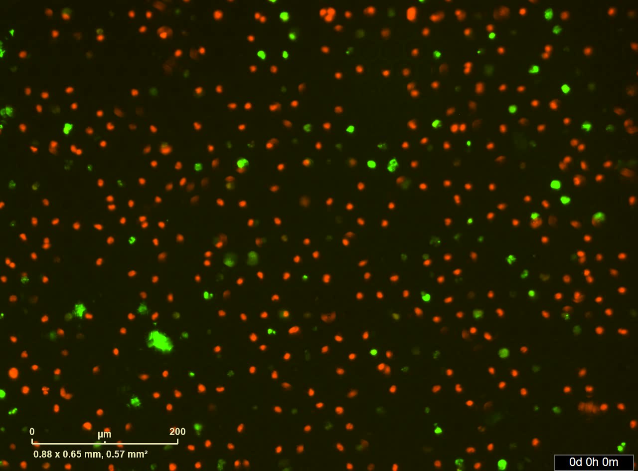
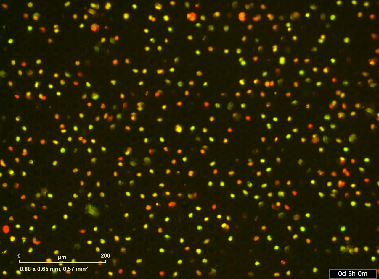
HL-60 cells were cultured for 3 days with 1.3% DMSO to induce differentiation. Neutrophil-differentiated HL-60 cells were incubated for 5 minutes with the membrane-permeable NUCLEAR-ID Red DNA dye to stain nuclei. After three times of washing, NUCLEAR-ID Red stained-neutrophils were loaded into SIEVEWELL. RPMI containing Sytox Green (0.2 µM) and 100 ng/mL PMA was added to induce NETosis and to assess for cell death. SIEVEWELL was placed on IncuCyte® S3 System which is housed inside a cell incubator at 37°C with 5% CO2. Neutrophils were imaged using phase contrast, red (800 ms exposure) and green (400 ms exposure) channels. Images using a 20x dry objective lens were taken every 5 minutes. >Live cell imaging
Reference of reagents, scan conditions. J Immunol January 15, 2018, 200 (2) 869-879.


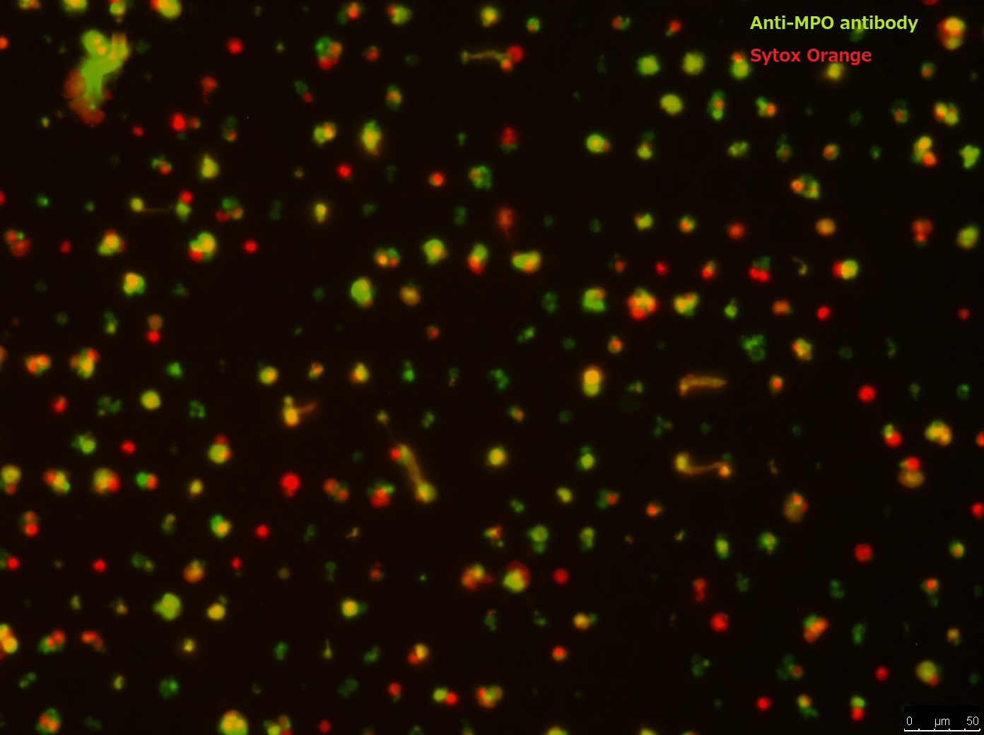
Human white blood cells, prepared from whole blood after RBC lysis, were loaded and incubated in SIEVEWELL with 100 ng/mL PMA containing RPMI medium for 4 hours to induce NETs. After washing out of medium, rabbit anti-MPO polyclonal antibody was added into SIEVEWELL and incubated for 30 minutes, then excess antibody was washed out. Alexa 488 labelled anti rabbit IgG and Sytox Orange was added and incubated for 30 minutes. Excess reagents were washed out from side ports and cells were washed with 2 mL PBS.

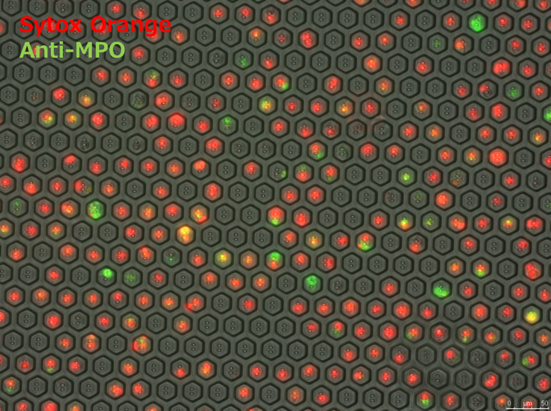
HL-60 cells were cultured for 3 days with 1.3% DMSO to induce differentiation. Neutrophil-differentiated HL-60 cells were loaded into SIEVEWELL and incubated directly in SIEVEWELL with 100 ng/mL PMA containing RPMI medium for 4 hours to induce NETs. After washing out of medium, rabbit anti-MPO polyclonal antibody was added into SIEVEWELL and incubated for 30 minutes, then excess antibody was washed out. Alexa 488 labelled anti rabbit IgG and Sytox Orange was added and incubated for 30 minutes. Excess reagents were washed out from side ports and cells were washed with 2 mL PBS.


