好中球細胞外トラップの検出
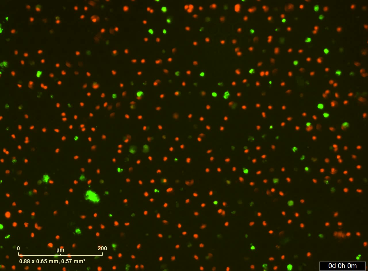
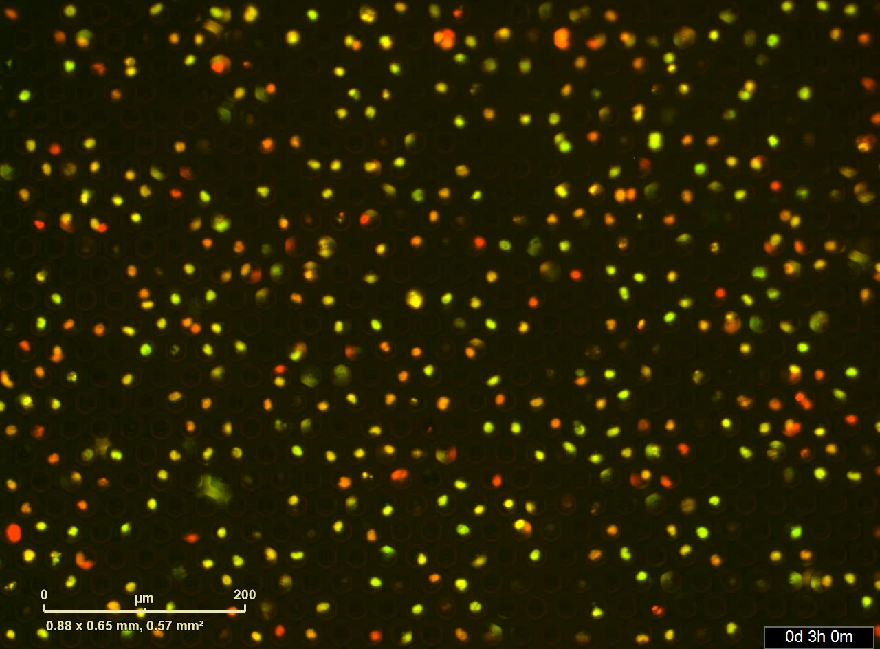
HL-60 ヒト前骨髄性白血病細胞株を1.3% DMSO添加RPMI1640培地で3日間培養し、分化誘導を行った。分化したHL-60細胞を細胞膜透過性色素であるNUCLEAR-ID Red DNA dyeで核を染色した。過剰の色素を洗浄除去した後、NUCLEAR-ID Redで染色した好中球をSIEVEWELLにロードした。Sytox Green (0.2 µM) と 100 ng/mL PMA を添加したRPMI培地を重層し NETosis を誘導した。細胞を播種したSIEVEWELLを IncuCyte® S3 システムに入れ、37°C 5% CO2 下で培養した。20倍レンズで5分ごとに位相差、赤 (800 ms 露光時間)、緑(400 ms 露光時間) の3チャネルで撮影した。>リアルタイムイメージング


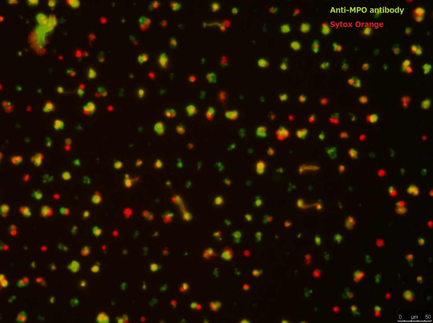
赤血球を溶血して得られたヒト白血球をSIEVEWELLに播種し、各ウェルに細胞を格納した。100 ng/mL PMAを含むRPMI培地で4時間培養し、NETsを誘導した。培地を吸引除去したのち、ウサギ抗MPOポリクローナル抗体を添加したPBSをロードし、30分間染色した。抗体を洗浄後、Alexa 488標識抗 ウサギIgG抗体およびSytox Orangeを添加したPBSをロードし、30分間染色した。染色後、未反応の試薬を洗浄により除去し、蛍光顕微鏡にて撮影した。

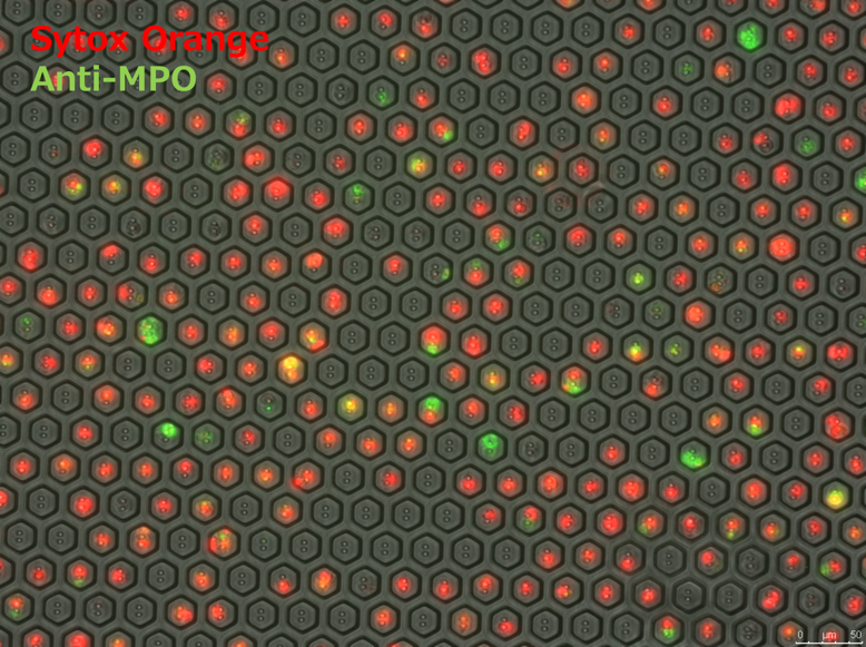
HL-60 ヒト前骨髄性白血病細胞株を1.3% DMSO添加RPMI1640培地で3日間培養し、好中球への分化誘導を行った。分化したHL-60細胞をSIEVEWELLに播種し、各ウェルに細胞を格納した。100 ng/mL PMAを含むRPMI培地で4時間培養し、NETsを誘導した。培地を吸引除去したのち、ウサギ抗MPOポリクローナル抗体を添加したPBSをロードし、30分間染色した。抗体を洗浄後、Alexa 488標識抗 ウサギIgG抗体およびSytox Orangeを添加したPBSをロードし、30分間染色した。染色後、未反応の試薬を洗浄により除去し、蛍光顕微鏡にて撮影した。


